
No products in the cart.
Return To Shop


Match a Multi-Modality Radiology Display to Modality Needs
A larger screen isn’t automatically the better radiology workstation. A multi-modality radiology display setup should match the imaging task, PACS workflow, GPU capacity, and available connectors; for primary mammography reading, a 12MP monitor is recommended, while a dual 5MP configuration is another option for mammography and tomosynthesis. The right arrangement also depends on how images and supporting applications need to appear together.
That balance matters: screen count and resolution shape how radiologists arrange studies, review findings, and move between worklists and reports. A display plan suited to one modality or reading task may not work for another.
This article explains how to match workstation layouts and display specifications to clinical use, assess graphics and connection requirements, and plan for mammography. It covers when a single large display, paired monitors, or separate non-diagnostic screens may fit, along with workflow checks to confirm the complete setup works as intended.
Key Takeaways
- Choose a single large display or multiple monitors based on the reading workflow, image arrangement, and need for separate applications.
- Match display resolution to the task, distinguishing primary diagnostic interpretation from clinical review and image acquisition.
- Assess GPU compatibility across monitor resolutions, connectors, bit depth, operating system, drivers, and PACS software.
- Plan mammography displays around the need to review breast images and tomosynthesis slices together, while keeping non-diagnostic screens separate from diagnostic use.
How do multimodality imaging workstations optimize the workflow?
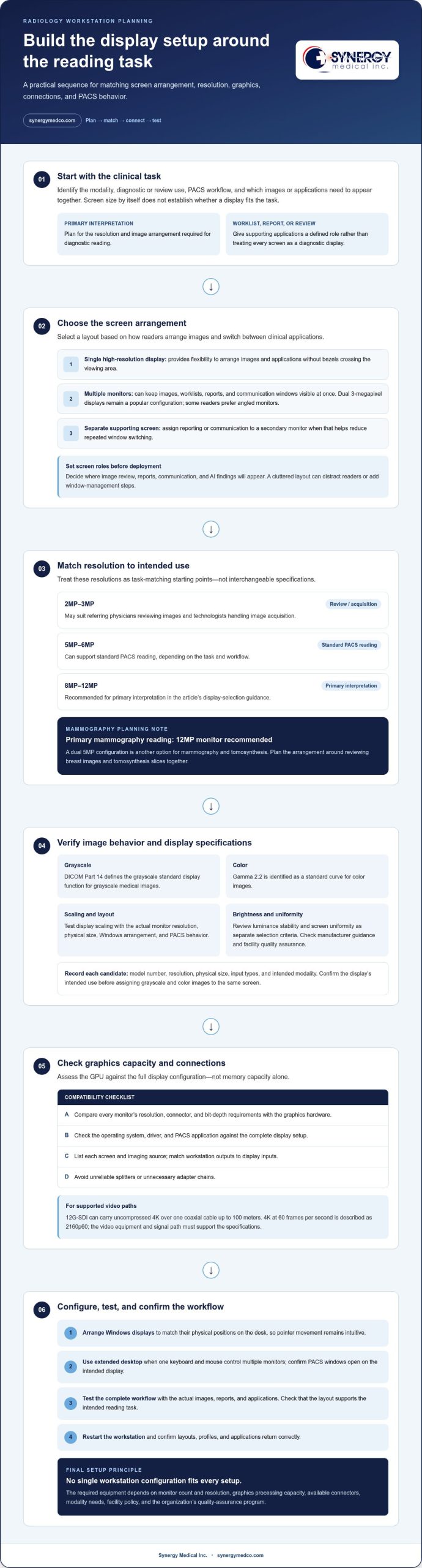
A single high-resolution display gives radiologists more freedom to arrange images and applications without bezels crossing the viewing area. A multi-monitor workstation can keep images, worklists, reports, and communication windows visible at once. The choice depends on modality, PACS workflow, and whether separate screens have distinct roles. Dual 3-megapixel displays remain a popular configuration, while some radiologists prefer angled monitors because the arrangement feels familiar.
One keyboard and mouse can control multiple monitors when the workstation is configured for an extended desktop. In Windows, arrange the displays to match their physical positions on the desk. This keeps pointer movement intuitive, such as moving from the primary image display to a report screen. Confirm that PACS windows open on the intended display.
Resolution should fit the imaging task, not just the screen size. For example, a display used for diagnostic interpretation has different demands from a screen showing a worklist or report. DICOM Part 14 defines the grayscale standard display function for grayscale medical images. Display scaling should be tested with the actual monitor resolution, physical size, Windows arrangement, and PACS behavior.
Assign each screen a role before deployment. A radiologist might use the primary display for image review and a secondary monitor for reporting or communication, reducing repeated window switching. Plan where AI findings appear, too: a cluttered layout can distract readers or add extra window-management steps.
In the United States, requirements depend on diagnostic use, facility policy, modality needs, and the organization’s quality-assurance program. Follow the monitor manufacturer’s guidance and the PACS vendor’s workflow when planning display quality assurance.
There is no single workstation configuration that fits every setup. The required equipment depends on the number and resolution of monitors, graphics processing capacity, available connectors, and modality needs.
Common deployment mistakes include mismatching the Windows display arrangement with the physical desk layout, copying profiles that do not suit every monitor, and failing to test PACS behavior. Test the complete workflow, then restart the workstation to confirm that layouts, profiles, and applications return correctly.
Choosing a display by model number, screen size, resolution, and inputs
Start with the model number, then compare the specifications that affect the intended reading task. Screen size alone does not show how much image detail a medical display can present. Radiology departments are showing interest in large-format displays with 6-megapixel, 8-megapixel, and 12-megapixel resolution. Dual 5-megapixel displays remain another established configuration for radiologists.
Match resolution to use. A 5MP or 6MP configuration can support standard PACS reading, while 8MP or 12MP displays are recommended for primary interpretation. Referring physicians reviewing images and technologists handling image acquisition may use 2MP or 3MP displays. These are starting points for matching the screen to the task, not interchangeable specifications.
For mixed-modality images, consider how grayscale and color studies will appear on the same screen. DICOM Part 14 defines the grayscale standard display function; gamma 2.2 is identified as a standard curve for color images. Confirm the display’s intended use and image-management workflow before assigning both image types to one screen. A single-screen configuration can reduce bezels and create room to arrange images and applications, but resolution and physical dimensions must support the required layout.
Check the inputs, too. Compare each display’s connectors with the workstation’s graphics outputs and the signals required by each source. Review the exact model specifications rather than assuming monitors with similar resolutions accept the same connections.
Treat luminance stability and screen uniformity as separate selection criteria. A backlight stabilization system can compensate for brightness changes related to temperature or aging. Review the manufacturer’s stated uniformity performance alongside the facility’s quality-assurance process. Consistent brightness across the screen supports reliable image review.
Before selection, record each candidate’s model number, resolution, physical size, input types, and intended modality. This comparison helps distinguish a suitable display from one that only matches the desk dimensions. Explore diagnostic monitor options when comparing display categories.
GPU capacity, connectors, and workstation compatibility
Assess the GPU against the complete display configuration, not memory capacity alone. Compare each monitor’s resolution, connector, and bit-depth requirements with the graphics hardware, operating system, driver, and PACS application. A workstation driving several high-resolution screens, for example, must support each at its intended resolution. GPU memory alone cannot establish compatibility.
List the connections required by each screen and imaging source before selecting a workstation. Match available outputs to monitor inputs, and avoid unreliable splitters or unnecessary adapter chains. For video workflows, 12G-SDI can carry uncompressed 4K over one coaxial cable up to 100 meters; 4K at 60 frames per second is described as 2160p60. These specifications apply when the video equipment and signal path support them.
Vendor-neutral workstations can bring mammography and other radiology applications into a shared clinical environment, provided every component works with the required software and displays. Diagnostic workstations support image interpretation; visualization workstations may also present images for review, collaboration, or planning. Keep the intended use clear: a screen used for worklists is not automatically suitable for diagnosis.
Configure PACS, RIS, and imaging modalities so each application opens on its assigned display and supports the facility’s reading workflow. For example, show PACS images on the diagnostic screen and RIS worklists or reports on a separate application display. Test modality image routing, application behavior, and display profiles together before clinical use.
Choose a workstation for its clinical role. A primary reader handling mammography needs graphics support for the diagnostic displays and software used in that workflow. A technologist’s acquisition station may have different display and application needs. Record monitor resolutions, connectors, operating system, driver, and PACS requirements, then assess the configuration as a complete system.
For a practical starting point, compare diagnostic display options against the workstation’s outputs and intended clinical tasks.
Mammography and multimodality display arrangements
Mammography reading requires an arrangement that supports comparison across views and studies. A 12MP display can present bilateral mammograms alongside digital breast tomosynthesis (DBT) slices, and a 12MP monitor is recommended for a primary mammography reading station. This can keep related images available together while readers assess the current study. The workstation’s display layout should preserve access to the images needed during interpretation.
A dual 5MP configuration is another option for mammography and tomosynthesis. The useful arrangement depends on how readers compare bilateral views, move through DBT slices, and refer to prior studies. Decide which images need simultaneous visibility, then map those views to the available screen area. Keep the display configuration consistent with the reading workflow and the PACS application’s image presentation.
Keep diagnostic reading separate from supporting tasks. Commercial displays may show worklists, reports, or communications, but do not treat them as diagnostic displays unless they are approved and managed for that use under organizational policies. For instance, keep a designated diagnostic screen focused on mammography images while another screen displays administrative applications.
Assess the planned arrangement using representative mammography studies before clinical use. Confirm that bilateral views and DBT slices are accessible in the intended sequence, and that the layout supports comparison without obscuring relevant images. Include the display manufacturer’s guidance, PACS behavior, modality requirements, facility policy, and the organization’s quality-assurance program in the evaluation.
The key planning decision is how the station will support primary mammography interpretation alongside other radiology tasks. Assign each screen a clear role and distinguish diagnostic reading from clinical review. This ties equipment selection to the actual reading task, not screen count alone, and gives the department a basis for evaluating proposed mammography and multimodality arrangements.
Synergy Medical Inc.: Surgical Monitors
Synergy Medical Inc. supplies Surgical Monitors, Radiology Monitors, and Mammography monitors for distinct clinical display needs. Use the workstation requirements above to identify the display category and resolution your department needs, then compare equipment suited to its intended role. Exam Lights are also available for clinical spaces where examination lighting is part of the equipment plan.
For a multi-modality radiology display setup, match the required diagnostic and mammography displays to the department’s workflow, workstation outputs, and clinical use. Synergy Medical Inc.’s monitor categories offer a practical starting point for selecting equipment.
Contact Synergy Medical Inc. to discuss monitor options.
Plan the Next Display Upgrade with Confidence
Plan displays as a shared clinical decision, not just a monitor purchase. Bring radiologists, imaging operations, and IT into the discussion so reading priorities, room constraints, and workstation compatibility are considered together. For a multi-modality radiology display setup, document each workstation’s purpose and who relies on it. This gives the department a clear reference when equipment or workflows change.
Before selecting equipment, align the clinical team on each display’s intended use and the operational team on system requirements. Shared planning can surface unresolved needs early, such as a workstation whose role has changed or a room that calls for a different arrangement. Synergy Medical Inc. offers Surgical Monitors, Radiology Monitors, and Mammography monitors for departments evaluating equipment for these needs.
A considered plan makes the next decision easier to communicate across clinical and technical teams. Start with your department’s specific needs.
With a clear plan, your team can move forward with confidence.
Frequently Asked Questions
What is a multimodality radiology display setup?
A multi-modality radiology display setup supports image review across modalities in a coordinated workstation environment. For example, a radiologist comparing a current study with prior imaging can arrange the studies side by side. Map these comparison tasks to the available viewing area and application behavior, so readers can move between studies without losing relevant context.
What monitors and hardware are needed for a radiology workstation?
A radiology workstation needs compatible displays, a computer, PACS access, and the input devices used by the reader. Before deployment, document the workstation’s users, applications, and peripheral needs. A shared reading station, for example, may need access configured for different staff roles, while a dedicated station can open directly into its primary reading workflow. This inventory reveals operational needs beyond display specifications.
What resolution and pixel density are recommended for diagnostic radiology displays?
Primary interpretation is recommended on 8MP or 12MP displays, while clinical review and image acquisition may use 2MP or 3MP displays. Pixel density also depends on the physical screen dimensions: the same resolution spread over a larger panel produces a different pixel density. Compare screen size and pixel count together, then assess whether the intended image arrangement remains legible at the workstation’s normal viewing distance.
What luminance, contrast ratio, and grayscale standards apply to radiology monitors?
Use the display manufacturer’s guidance and the facility’s quality-assurance process to assess luminance and grayscale performance. DICOM Part 14 defines the grayscale display function for medical images. Contrast ratio is a separate specification and should not be treated as a substitute for grayscale response. Review both specifications in context, including the display’s intended role and how its images appear in the workstation’s normal viewing environment.
How do diagnostic displays differ from clinical review displays?
A diagnostic display is designated for interpretation, while a clinical review display supports other image-viewing tasks. Make each screen’s role clear to staff with workstation labels or documented room procedures. For example, a clinician reviewing images during a consultation should be able to distinguish that screen from the one assigned to primary interpretation. This helps prevent a convenience screen from being mistaken for an approved diagnostic display.
What are the DICOM GSDF and AAPM TG18 requirements for radiology displays?
DICOM Part 14 defines the Grayscale Standard Display Function, or GSDF, for grayscale medical images. GSDF describes grayscale presentation, not resolution, connector type, or pixel density. Apply the display manufacturer’s guidance and the facility’s quality-assurance program when assessing display performance. Keep calibration and QA records with the workstation documentation so the team can refer to the display’s assigned profile and evaluation history.
About author
